DNA Mutation

When observing the frequency of mutations of one particular gene sequence across a population or species, it would be very uncommon to find a mutation. If one were to look at an entire human genome that contains millions of amino acid sequences, one will find that they are quite common on that scale. The frequency at which a mutation will occur has a positive correlation to the genome size. A mutation is simply a change in DNA that can occur randomly but could be caused by certain genetic or environmental factors. It is important to understand that a change in DNA is only considered a mutation when the change in DNA makes it past the repair and regulatory mechanisms. When we say mutations occur at random we mean they occur without any regard to the needs of the organism in question.Though most tend to see Mutations as only having a negative impact on life, they are one of the primary driving forces behind genetic variation, speciation, and evolution. Most mutations originate from somatic cells, which are continuously dividing and are therefore more susceptible to mutation. Some examples include skin, mucous membranes, muscle and skeletal tissues. Luckily these mutations stay within the individual and are not passed on to progeny. Luckily Only Germ-line mutations can be passed down to progeny in mammals. Some somatic mutations in plants can be passed down to progeny as long as the plant produces viable offspring.

There are many different kinds of mutation that can affect a genome on both small and large scales. The small scale mutations include point mutations, insertions and deletions, and transposable elements. They are considered small because they normally only affect one particular nucleotide base pair or protein (if anything at all). Point mutations are changes to a single nucleotide within a gene. In most cases (with humans), the mutation goes unnoticed without any phenotypic changes because the majority of codons in our genome have no known function. Sometimes the base pair does not even change. A point mutation that does not change a nucleotide base pair is a synonymous (aka silent) Mutation. One that does change the sequence is called a nonsynonymous mutation and may or may not have any detrimental effects depending on where the mutation occurred and if it encodes for a protein. A nonsense mutation is of the more dangerous kinds. They are mutations that cause an amino acid sequence to change, resulting in a STOP codon to appear when it should not. If the sequence encodes for a protein, it will be defective. Transposable elements are amino acid sequences that can insert themselves into an open sequence and are also expected to negatively impact gene expression or translation.

Insertions and deletions are particularly dangerous because inserting an unnecessary base pair into a strand of DNA or deleting a sequence that should be there will cause changes in the genome all the way downstream, resulting in loss of function or alteration of proteins. Normally a cell will not survive loss-of-function mutations of this type.

The large scale mutations include gene duplications, inversions, nondisjunction, reciprocal translocations etc. A duplication occurs when a whole region of a chromosome is present two times, and a deletion is when part of a chromosome is not present at all. Reciprocal translocations occur when two segments switch with each other and can eventually lead to complications during recombination in meiosis. Lastly, inversions occur when a chromosome segment is inserted opposite to how it is supposed to be.

Overall, Mutations aid in genetic diversity, but can also be dangerous. Luckily the most dangerous mutations such ad deletions, nonsense, and frameshift mutations are not passed down to progeny because the cell or organism in question probably wont live that long.

Biology: How life works (2nd ed.). (2017). In B. A. Berry Andrew, Biology: How life works (2nd ed.) (p. CH 14). McGraw Hill, NY: W. H. Freeman. Retrieved from http://www.macmillanhighered.com/launchpad/morris2e/4909413#/ebook/item/MODULE_bsi__F4951CED__2971__4486__BC4F__E109B2EE87D4/bsi__2ED633B9__F1E8__4C6C__82AA__0633C0C9F474?mode=Preview&toc=syllabusfilter&readOnly=False&renderIn=fne

Molecular Phylogeny

Molecular Phylogeny

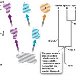
Molecular phylogeny is a method used to analyze molecular differences in heritable traits within a gene. These traits are compared to other organisms that are thought to be genetically similar on order to determine whether there is an evolutionary relationship between them. Phylogenetic trees can give us a bigger picture of the relationships between species, if they have common ancestors, or if the species or phylogenic groups can be traced back to in the phylogenic tree to a point where we can hypothesize a general time frame when one subset of a species genetically diverged from an original. It will be important to remember that a species includes organisms able to interbreed and produce viable offspring. When imagining the origins of life, we can see how putting together all evolutionary relationships would form a tree, with the most shared common ancestors at the base and trunk, and the most recent species forming the branches. Below is an example of a phylogenetic tree retrieved from Chapter 23 of Launchpad:

Looking above, one can see that the origin of the tree in question is called the “root” named so because that is the most common ancestor of species A, B, and C. the forks where we can see genetic divergence is called a node. The lines coming off of that node is called a Branch, which can be followed to yet another divergent species. The more branches observed in a phylogenetic diagram, the more speciation that occurred.

References

Biology: How life works (2nd ed.). (2017). In B. A. Berry Andrew, Biology: How life works (2nd ed.) (p. CH 23). McGraw Hill, NY: W. H. Freeman. Retrieved from http://www.macmillanhighered.com/launchpad/morris2e/4909413#/ebook/item/MODULE_bsi__F4951CED__2971__4486__BC4F__E109B2EE87D4/bsi__2ED633B9__F1E8__4C6C__82AA__0633C0C9F474?mode=Preview&toc=syllabusfilter&readOnly=False&renderIn=fne
Enzymes & Enzyme Inhibitors

Enzymes play many key roles in energy metabolism and homeostatic balance. Without them, cells would be several times more inefficient (if not nonfunctional) in its energy usage, more susceptible to foreign materials, and wouldn’t be able to utilize as many organic materials for energy. Enzymes are proteins. Like all proteins, they are made up of long chains of amino acids transcribed from DNA or RNA templates. They catalyze/accelerate the rate of chemical reactions by lowering the activation energy needed to carry out said reaction. Activation energy is the input of energy required by all chemical reactions to break/synthesize bonds for metabolism in an efficient manner. They accomplish this by stabilizing reactant(s) in their transition state, lowering the amount of free energy released to make it more available, lowering the amount of input energy needed to make or break a chemical bond. Enzymes are also energy efficient because they do not disappear after use and can continue to catalyze reactions. There are several different kinds of enzymes, and each type works on a substrate or reaction. Their specificity comes from the fact that the shape of the enzyme will determine its overall function, so therefore they are tailored to work on one substrate.

All enzymes have special folds in its structure designed to fit a specific substrate, called an Active site. Once a substrate is attached to the active site, a cofactor is normally needed in another receptor or fold of the enzyme for it to perform its function. Cofactors can include Vitamins, co-enzymes (enzymes that work in conjunction with another to form a complex), metal ions, hormones, etc.
Though most enzymes are regulated by cell processes like gene expression, hormone and neurotransmitter release (forms of cell signaling) negative feedback loop with its substrate, or cofactor availability, enzymes are greatly affected by their environment. Its shape can be altered in the presence charged molecules, very susceptible to changes in PH and light, and can be inhibited. Enzyme inhibition is a useful concept to know because inhibitors are used a lot in the medical field in drug therapy, in slowing an unwanted reaction, etc. A most well-known example is penicillin, which inhibits an enzyme used by pathogenic bacteria that repairs damage to its cell wall.

There are three types of enzymatic inhibition: Competitive inhibition, noncompetitive inhibition, and allosteric inhibition. Competitive inhibition occurs when a molecule similar in structure to the substrate binds to its active site, thus preventing it from performing its function (penicillin is an example). Noncompetitive inhibition is when an inhibitor binds to an area other than its active site. They could work by changing its shape or possibly by physically blocking the active site. Lastly, allosteric inhibitors work by adding either an activator molecule to its allosteric site (the site where activators/inhibitors bind to). Examples of allosteric regulators could be hormones, neurotransmitters, or some other type of cell signaling molecule.

References

Biology: How life works (2nd ed.). (2017). In B. A. Berry Andrew, Biology: How life works (2nd ed.) (p. CH 6). McGraw Hill, NY: W. H. Freeman. Retrieved from http://www.macmillanhighered.com/launchpad/morris2e/4909413#/ebook/item/MODULE_bsi__F4951CED__2971__4486__BC4F__E109B2EE87D4/bsi__2ED633B9__F1E8__4C6C__82AA__0633C0C9F474?mode=Preview&toc=syllabusfilter&readOnly=False&renderIn=fne
Hardy-Weinberg Principal

The Hardy-Weinberg principle is a concept conceived in 1908 by G.H Hardy and Wilhelm Weinberg. It describes a situation in which allelic or genotypic frequencies that drive evolution do not change over time in a population, and therefore that population is not evolving. The concept is based on Gregor Mendel’s law of independent assortment, which states that alleles of a trait can be separated independently from each other and can lead to fluctuations in the frequency of alleles over time. With this fact known, Hardy and Weinberg developed a series of conditions that must be met in order to determine whether or not a population is evolving. This established baseline is very useful in the study of population genetics. The conditions that must be met are as follows:

1. There can be no differences in the survival and reproductive success of individuals in the population. If a certain genotype of individuals in a population have a better chance of survival than the others or if a homozygous recessive gene expresses for some deadly condition, then we know automatically that nature will select for the genotype most fit to its environment and evolution would then occur.

2. Populations must not be added to or subtracted from by migration. If one has a population they are studying and added or subtracted individuals, the frequency of the gene(s) in question being observed would immediately change because the proportion of alleles to individuals would be thrown off. Because evolution is defined as a change in allele frequency over time in a population, a change in frequency by the addition or subtraction of that genotype would count as a population evolving.

3. There can be no mutation occurring. Simply put, mutations are one of the ways populations can evolve. It should be of note to mention that mutation is not the biggest concern because population geneticists generally study populations in a much shorter timescale than it would take for mutations to accumulate. Still, none allowed.

4. The population must be large enough to prevent sampling errors. A sampling error occurs when not enough specimens in that population are being observed. If the population is too small, then those individuals have a much bigger impact on allele frequency than one would see in an actual population and any statistical data gained could not possibly reflect the population as a whole, leading to inaccurate data. A smaller population would also be more impacted by genetic drift, which is also a form of evolution.

5. Matings within the population must be at random and not chosen or planned. If individuals with known allele frequencies were chosen to mate, It would interfere with your results, so mating must happen without any regard to phenotype, as would be the case in nature. This doesn’t affect the overall genotype frequency but not the frequency of alleles (because of independent assortment).

This test can have big applications in population genetics.On top of determining whether or not a population is/has evolved, we can also use it to make predictions on what evolutionary factors are affecting a population, predict allele frequencies in a population, and use it to look for correlations between genotype frequency and allele frequency.

References

Biology: How life works (2nd ed.). (2017). In B. A. Berry Andrew, Biology: How life works (2nd ed.) (p. CH 21). McGraw-Hill, NY: W. H. Freeman. Retrieved from http://www.macmillanhighered.com/launchpad/morris2e/4909413#/ebook/item/MODULE_bsi__F4951CED__2971__4486__BC4F__E109B2EE87D4/bsi__2ED633B9__F1E8__4C6C__82AA__0633C0C9F474?mode=Preview&toc=syllabusfilter&readOnly=False&renderIn=fne

Cytokinesis

Cytokinesis is the stage in the eukaryotic cell cycle where a replicating cell divides its components into 2 separate membranes, forming daughter cells. It is widely thought that cytokinesis in eukaryotes evolved from the similar process of binary fission in Prokaryotes, yet it is a more complex process in comparison due to the nature of eukaryotic DNA replication. Cytokinesis is also more complex because eukaryotes have nuclei that must break down in the beginning of the cell cycle and reform at its end. It occurs in Both meiosis and mitosis wherein in the final stage of mitosis creates 2 diploid daughter cells with the same genetic makeup as the parent cell, and in meiosis (the reproduction of germ-line cells) it occurs twice. the first division occurs after telophase of Meiosis I, and again in Meiosis II, resulting in 4 unique haploid cells. The cells are haploid in meiosis because each daughter cell contains only one pair of chromosomes that will later recombine with another germ-cell of the opposite sex.

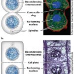
The Picture shown above retrieved from the Biology Launchpad website shows cytokinesis in both plants and animals. All eukaryotes perform cytokinesis to divide genetic material and cytoplasmic components amongst the daughter cells, but the mechanism used to perform the separation varies between plants, animals, and fungi. In mammals, a contractile ring made of actin filaments forms at the center of the cell, and motor proteins cause the filaments to contract and will eventually pinch the cell in two, resulting in 2 new daughter cells. Plants are different because they have cell walls and do not physically separate from each other. Once the plant cell doubles in size it will create a wall of microtubules in the center called a pragmoplast that will pull in vesicles containing cell wall components towards it during telophase. The vesicles will then fuse at the center (or cell plate) to form a cell wall that is attached to the original. Fungal cytokinesis is more like animal cytokinesis and uses a contractile ring made of actomyosin.

References

Biology: How life works (2nd ed.). (2017). In B. A. Berry Andrew, Biology: How life works (2nd ed.) (p. CH 11). McGraw-Hill, NY: W. H. Freeman. Retrieved from http://www.macmillanhighered.com/launchpad/morris2e/4909413#/ebook/item/MODULE_bsi__F4951CED__2971__4486__BC4F__E109B2EE87D4/bsi__2ED633B9__F1E8__4C6C__82AA__0633C0C9F474?mode=Preview&toc=syllabusfilter&readOnly=False&renderIn=fne

“The Mitotic Phase and the G0 Phase.” Boundless Biology Boundless, 24 Mar. 2017. Retrieved 24 Apr. 2017 from https://www.boundless.com/biology/textbooks/boundless-biology-textbook/cell-reproduction-10/the-cell-cycle-88/the-mitotic-phase-and-the-g0-phase-396-11622/

ATP

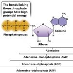
All cells require ATP to perform any action where an input of energy is needed. ATP stands for Adenosine triphosphate and is well known as the “energy currency” of all cells. We relate it to currency because ATP is not the energy itself, rather it is a molecule with a high energy value stored in its chemical bonds. Its role in metabolism is similar to the way a dollar bill represents purchasing power. The more ATP a cell contains, the more energy it has readily available for use when needed. ATP is composed of a triphosphate group (a chain of three phosphates) and adenosine (an adenine attached to a five-carbon ribose sugar). Below is a picture representing its chemical structure.

Because its energy value comes from its chemical bonds, ATP is considered a form of chemical potential energy. The reason it has such a high energy value relative to other molecules is primarily because of its unstable phosphate group. The bonds are unstable because phosphates are negatively charged and try to repel each other, but they are held together by weak hydrogen bonds. The fact that the phosphates are naturally repelling each other due to their opposing charges and are held together by weak hydrogen bonds make it easy for a cell to break the bond in a hydrolysis reaction, and in doing so will transform its stored chemical energy into mechanical energy that will be used to drive most cellular processes.
Though one molecule of ATP is structurally identical to others, the energy output from the hydrolysis of ATP can vary. The maximum output from the hydrolysis of ATP within a living cell can be up to -57kj/mol, however environmental conditions within the cell like PH and Magnesium ion (Mg2+) concentration can change this value. Note the negative sign implies energy is released from the reaction, so it is classified as exergonic. The second law of thermodynamics states that energy will be inevitably lost in the reaction in the form of heat, so it is impossible for the reaction to be 100% efficient. To compensate for this loss ATP catabolisms are coupled with chemical reactions of other forms, namely with redox reactions. The synthesis reaction of ATP in the form of a chemical equation is ADP+Pi+free energy–>ATP + H2O. ADP stands for Adenosine diphosphate and is a more stable molecule. This reaction is reversible, and therefore adding a Pi group to ADP with enough added energy will yield ATP. Clearly, without ATP, life on earth would not be possible, so understanding the role of ATP in energy metabolism is critical to understand life’s processes.

References

ATP: Adenosine Triphosphate. (2017). Retrieved April 24, 2017, from Boundless Biology Boundless: Retrieved from https://www.boundless.com/biology/textbooks/boundless-biology-textbook/metabolism-6/atp-adenosine-triphosphate-71/atp-adenosine-triphosphate-349-12938/

Biology: How life works (2nd ed.). (2017). In B. A. Berry Andrew, Biology: How life works (2nd ed.) (p.115-123, CH 6). McGraw-Hill, NY: W. H. Freeman

Semipermeable vs Selectively permeable Membranes

Semipermeable vs Selectively Permeable Membranes

The term permeability in biology always refers to membranes. These membranes are made of lipids (phospholipids and cholesterol), integral and peripheral proteins, and carbohydrates (glycolipids and glycoproteins) that all interact with each other to form a barrier between the cell and its environment. The proportion of Carbohydrates, lipids, and fats in membranes vary by cell type and species, but in humans, they are about 50% protein, 40% lipid, and 10% carbohydrates. Cell Membranes are fluid (dynamic in movement) and can regenerate up to a certain degree when damaged. More importantly, cell membranes maintain the electrochemical gradient between the inside of a cell and its environment and can allow smaller charged molecules, water, and metabolic waste to pass in and out of it, making them permeable. This permeability is therefore a vital aspect in maintaining homeostasis.
When referring to membrane permeability there are two types found in living things: semi-permeable and selectively permeable. Both allow molecules and water to move in and out of the cell, as needed to maintain homeostasis. Semipermeable membranes are more simple in function because they are not “picky”, so If molecules are small enough they will pass through the membrane by osmosis, diffusion or following its concentration gradient from an area of higher concentration to an area of lower solute concentration. One example of a semipermeable membrane found in the body would be the tubules of nephrons within the kidney. Blood components like red blood cells, large proteins that are too large to pass through the nephrons will not pass through the tubules, while smaller solutes, Na+, and metabolic waste passes through the kidney to ultimately become filtrate in urine. Patients with renal problems who can’t properly filter blood must undergo dialysis, where an external synthetic semipermeable filter that acts as a membrane is used much like functional kidneys would.
Selectively permeable membranes are more specific (hence the name selective) as to what passes through the membrane, and when. Cell membranes are considered selectively permeable; Some molecules like water can freely pass in and out to regulate solute concentration within the cell, other molecules such as Sodium (Na+), Potassium (K+), carbon dioxide (CO2), hormones and growth factors are regulated. Of course, some molecules are not allowed in at all. Particles that are needed by the cell but cannot diffuse through the membrane on its own can pass through via active transport with the help of integral proteins permanently integrated in the cell wall, and by transport proteins that carry the molecule to wherever it needs to go to be broken down and utilized. The membrane also has pumps that use ATP to expel solutes like Na+ and K+ out of the cell, and receptors (or ligands) that allow for the passage of larger solutes into it.
Though plasma membranes in cells let some molecules like water and sodium pass through freely, they cannot be considered semipermeable because they have a degree of control over what goes in and out to maintain homeostasis. Regulation of this degree can only be done by selectively permeable membranes, and without being selective of what can pass through it the cell would not be able to maintain its inner environment and eventually die.

Semipermeable vs selectively permeable membranes-1e3kmwf

Types of RNA

In living organisms, RNA (ribonucleic acid) is transcribed from DNA (deoxyribonucleic acid). They are later transcribed to be expressed into proteins that are needed for structure and repair, metabolic functions, and regulations to include defense. In comparison to DNA, RNA transcripts are single-stranded, shorter in length, contain a ribose sugar backbone instead of Deoxyribose, and use the nitrogen base uracil instead of thymine. RNA is also less stable than DNA and are therefore more susceptible to reacting with its environment. There are 5 different types of RNA that all perform specific functions that relate to protein expression or regulation that include: messenger RNA (mRNA), transfer RNA (TRNA), ribosomal RNA (rRNA), small nuclear RNA (snRNA), and micro RNA (miRNA). Not all types are present in every domain of life, and the amount and rate in which they are produced varies per the needs of the cell.

mRNA is processed from the primary DNA transcript in bacteria and eukaryotes. Their main function after its transcription is to carry the protein encoding message to ribosomes (composed of 60% rRNA and 40% protein) to be translated. They are read in three sets of triplet nucleotide pairs called codons in which three consecutive codons will translate for one protein. In eukaryotes, mRNA is transcribed within the nucleus and exit through nuclear pores to be translated by ribosomes. Prokaryotes do not have nuclei, and its transcription is coupled with its translation while it separates from the primary transcript. Prokaryotic mRNA is also different in that one strand can be polycistronic, or can code for more than one protein. Eukaryotic mRNA must go through RNA processing for translation. Alternatively, eukaryotic mRNA that encodes for one gene can be modified by alternative splicing (removing of introns) to change how it is expressed.

Around 80% of the genes in humans are alternatively spliced. This splicing is done by ribonucleoprotein complexes called spliceosomes which are made of snRNA or scRNA added to proteins. snRNA is present in eukaryotes, whereas scRNA (small cytoplasmic RNA) is found in both prokaryotes and eukaryotes. Some types of scRNA are involved in post-translational modification of proteins that can regulate the protein after its made, or can activate or inactivate them.

rRNA is present in all living cells and is one of the most abundant in mammals. Its primary function is to use the mRNA template to translate an encoded gene and synthesize it into a protein. In both eukaryotes and prokaryotes, the ribosomes are present in the cytoplasm. The rRNA complex within these ribosomes are composed of 1-3 subunits composed of a large subunit (LSU) and up to 2 small subunit (SSU), where eukaryotic ribosomes are larger. Though ribosomes are the site of protein synthesis and establish the right reading frame, it is tRNA that performs the synthesis of the mRNA strand into a functional protein.

tRNA are molecules located within the ribosome. They are composed of nucleotide chains ranging from 70-90 in length. The tRNA carries free-floating amino acids within the cytoplasm along these chains, and “transfer” them to the mRNA-rRNA complex. Attachment of the amino acid to uncharged tRNA is done by an enzyme called aminoacetyl tRNA synthase. Each type of aminoacetyl tRNA synthase enzyme works on only one of the 20 amino acids. Once attached, the tRNA is considered “charged” and the amino acid is attached at the 3’ end. will then travel to the reading frame in the ribosome to the mRNA to be added at its 5’ end in an antiparallel configuration, and therefore are referred to as anticodons.

In conclusion, all types of RNA are involved in some step of protein synthesis. Some types are specific by the type of organism in question, while others (such as rRNA) are present in all living organisms, most likely because RNA evolved over time to make DNA and similarities of RNA can be traced back to life’s origins.

References

Nester’s Microbiology: a human perspective (8th ed.. (2015). ch 4 In A. D. Allen Debra. McGraw Hill, NY: McGraw-hill.
Openstax. (2013). Biology. In Openstax, Biology (p. Ch1.1). Houston, TX: Rice University.

Phospholipids

Image

Phospholipids

Lipids are a class of macromolecules that are vital to life and have special properties that aid in the structure, maintenance, and metabolic activities of living organisms. Their exact origin has not yet been discovered, however it is clear that lipids were formed by straightforward processes in the environment, and over time they eventually developed into more complex structures (like the many varieties of phospholipids) that form membrane components of cells. Because they can form membranes, lipids must have been to be one of the first processes that started life to occur because cells could never survive without a protective barrier protecting and regulating their internal components from its respective environment. The most important properties of lipids are their polar nature, making them amphipathic (meaning they have hydrophilic and hydrophobic parts). Phospholipids are macromolecules that are made up of a phosphate head group, glycerol backbone, and 2 chains of fatty acid tails. Below is a photo showing their structure and the variety of ways they can be represented:

https://sites.gsu.edu/biol2107teamnucleus_maxwell/wp-admin/media-upload.php?post_id=33&type=image&TB_iframe=1
(Photo retrieved from Biology: How life works, 2nd edition)

Their amphipathic nature is what gives phospholipids their ability to form membranes. The polar head groups are hydrophilic and make them “water loving”, and their hydrophobic tails are hydrophobic, or “water fearing”. This becomes apparent when enough phospholipids are added to an aqueous solution. The Polar heads will attach to each other by Van der waals forces (the association between molecules by their slight charges), and the fatty acid tails would will line up side-by-side. Because the polar tails want to be separated from water, the polar heads will eventually form a protective barrier to the aqueous environment. The varying bulk of the polar heads will determine what kind of membrane will form. They can make spherical micelles that have the nonpolar tails oriented on the inside, a fluid bilayer that has the tails sandwiched inside 2 layers of polar heads, and spherical vesicles that have a layer of polar heads both inside and outside of the cell.
The fact that these lipids spontaneously come together in the previously mentioned fashion gives them an ability to repair itself when the phospholipid bilayer is broken. If phospholipids from the environment or made from cellular processes are present, they will be added to the membrane until the damage is repaired. This along with its amphipathic nature makes them very effective membranes. Phospholipids can also associate with cholesterol, proteins, and other slightly charged molecules needed for metabolism to become selectively permeable. This is shown in all cell membranes because molecules needed for energy consumption and regulation must be able to enter and exit the cell as needed to maintain a proper electrochemical gradient, PH, and solute concentration if the cell is to survive.

References
Biology: How life works (2nd ed.). (2017). In B. A. Berry Andrew, Biology: How life works (2nd ed.) (p. CH 5.1). McGraw Hill, NY: W. H. Freeman. Retrieved from http://www.macmillanhighered.com/launchpad/morris2e/4909413#/ebook/item/MODULE_bsi__F4951CED__2971__4486__BC4F__E109B2EE87D4/bsi__2ED633B9__F1E8__4C6C__82AA__0633C0C9F474?mode=Preview&toc=syllabusfilter&readOnly=False&renderIn=fne
Openstax. (2013). Biology. In Openstax, Biology (p. Ch1.1). Houston, TX: Rice University.
Suetsugu, S. K. (2014). Dynamic shaping of cellular membranes by phospholipids and membrane-deforming proteins. Physiological Reviews, 94(4), 1219-1248. Retrieved April 3, 2017, from http://physrev.physiology.org/content/94/4/1219