Final Learning Summation Pt. II

Oxidative Phosphorylation

Oxidative Phosphorylation is a process where energy is sustained throughout a series of protein complexes that occurs in the inner-membrane of the mitochondria which ends up making ATP. This process is broken down into two parts, oxidation of NADH and FADH2 and Phosphorylation. In the first step, both NADH and FADH3 both experience the process of losing electrons through oxidation. Both NADH and FADH3 will transfer their high energy molecules into two different protein complexes (Protein Complex I and Protein Complex II). The Oxidation of NADH results to a pumping of protons through Protein Complex I. The electrons that were received by Protein Complex I are then given to an electron-carrier called Ubiquinone (Q). On the other hand, FADH3 goes through a similar process that NADH goes through when these two high energy molecules goes through with oxidation. Afterwards, an electrochemical gradient has been created, meaning both sides are different in electrical charge. The protons on the outside of mitochondrial membrane will then push through the ATP synthease. This movement of protons causes ATP synthase to spin, and bind ADP and Pi, producing ATP.

PCR

Polymerase Chain Reaction (PCR) is a laboratory technique used to make millions of copies of a specific region of DNA. This technique can be used for research on a specific gene that may hold a recessive trait or finding the genetic marker in the forensic science field during a crime investigation. Typically, the goal of PCR is to make enough of the target DNA region that it can be analyzed or used in some other way. For instance, DNA amplified by PCR may be sent for sequencing, visualized by gel electrophoresis, or cloned into a plasmid for further experiments. PCR is used in many areas of biology and medicine, including molecular biology research, medical diagnostics, and even some branches of ecology.

Control of Cell Cycle

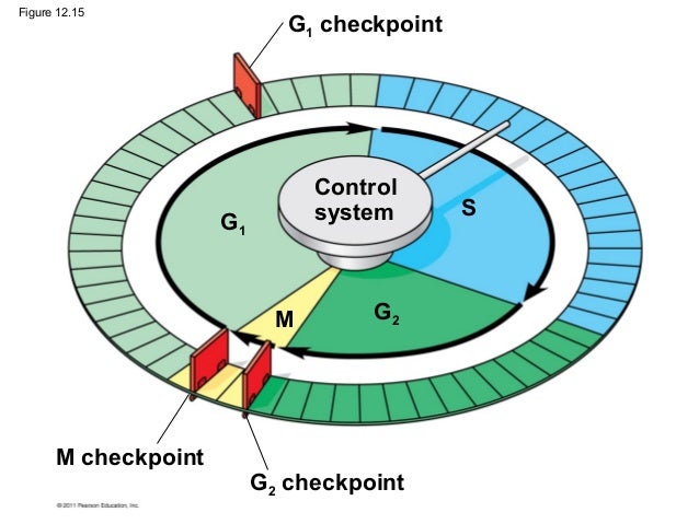
The Cell Cylce control cycle works just like the control system on a washing machine, and as the cell goes through with DNA replication, mitosis and etc, the process is controlled by a system. There are four stages that the cell cycle is controlled by which are G1, G2, S, and M phase. The control system is effected by either internal or external factors. At each check point, the cell cycle will stop and then proceed to the next cycle when there is a given signal to proceed.

Image result for cell cycle control system

References:

https://www.khanacademy.org/test-prep/mcat/biomolecules/krebs-citric-acid-cycle-and-oxidative-phosphorylation/a/oxidative-phosphorylation-the-major-energy-provider-of-the-cell

https://www.khanacademy.org/science/biology/biotech-dna-technology/dna-sequencing-pcr-electrophoresis/a/polymerase-chain-reaction-pcr

https://www.ncbi.nlm.nih.gov/books/NBK26824/

DNA Mutation

When observing the frequency of mutations of one particular gene sequence across a population or species, it would be very uncommon to find a mutation. If one were to look at an entire human genome that contains millions of amino acid sequences, one will find that they are quite common on that scale. The frequency at which a mutation will occur has a positive correlation to the genome size. A mutation is simply a change in DNA that can occur randomly but could be caused by certain genetic or environmental factors. It is important to understand that a change in DNA is only considered a mutation when the change in DNA makes it past the repair and regulatory mechanisms. When we say mutations occur at random we mean they occur without any regard to the needs of the organism in question.Though most tend to see Mutations as only having a negative impact on life, they are one of the primary driving forces behind genetic variation, speciation, and evolution. Most mutations originate from somatic cells, which are continuously dividing and are therefore more susceptible to mutation. Some examples include skin, mucous membranes, muscle and skeletal tissues. Luckily these mutations stay within the individual and are not passed on to progeny. Luckily Only Germ-line mutations can be passed down to progeny in mammals. Some somatic mutations in plants can be passed down to progeny as long as the plant produces viable offspring.

There are many different kinds of mutation that can affect a genome on both small and large scales. The small scale mutations include point mutations, insertions and deletions, and transposable elements. They are considered small because they normally only affect one particular nucleotide base pair or protein (if anything at all). Point mutations are changes to a single nucleotide within a gene. In most cases (with humans), the mutation goes unnoticed without any phenotypic changes because the majority of codons in our genome have no known function. Sometimes the base pair does not even change. A point mutation that does not change a nucleotide base pair is a synonymous (aka silent) Mutation. One that does change the sequence is called a nonsynonymous mutation and may or may not have any detrimental effects depending on where the mutation occurred and if it encodes for a protein. A nonsense mutation is of the more dangerous kinds. They are mutations that cause an amino acid sequence to change, resulting in a STOP codon to appear when it should not. If the sequence encodes for a protein, it will be defective. Transposable elements are amino acid sequences that can insert themselves into an open sequence and are also expected to negatively impact gene expression or translation.

Insertions and deletions are particularly dangerous because inserting an unnecessary base pair into a strand of DNA or deleting a sequence that should be there will cause changes in the genome all the way downstream, resulting in loss of function or alteration of proteins. Normally a cell will not survive loss-of-function mutations of this type.

The large scale mutations include gene duplications, inversions, nondisjunction, reciprocal translocations etc. A duplication occurs when a whole region of a chromosome is present two times, and a deletion is when part of a chromosome is not present at all. Reciprocal translocations occur when two segments switch with each other and can eventually lead to complications during recombination in meiosis. Lastly, inversions occur when a chromosome segment is inserted opposite to how it is supposed to be.

Overall, Mutations aid in genetic diversity, but can also be dangerous. Luckily the most dangerous mutations such ad deletions, nonsense, and frameshift mutations are not passed down to progeny because the cell or organism in question probably wont live that long.

Biology: How life works (2nd ed.). (2017). In B. A. Berry Andrew, Biology: How life works (2nd ed.) (p. CH 14). McGraw Hill, NY: W. H. Freeman. Retrieved from http://www.macmillanhighered.com/launchpad/morris2e/4909413#/ebook/item/MODULE_bsi__F4951CED__2971__4486__BC4F__E109B2EE87D4/bsi__2ED633B9__F1E8__4C6C__82AA__0633C0C9F474?mode=Preview&toc=syllabusfilter&readOnly=False&renderIn=fne

Final Learning Summation pt. I (Last Lecture Day)

Cellular Energetics

In photosynthesis, energy enters as a form of light which will then be converted into chemical energy, and carbon dioxide and water are used to store the energy in the form of carbohydrates. The carbohydrate is then taken apart in respiration and the chemical energy is transferred to a different compound called ATP (provides the organelles to do work). In chemosynthesis, energy enters the system in the form of inorganic compounds which have stored energy. The energy is transferred to the bonds of a carbohydrate, meaning that the remaining reactions are the same as a photosynthetic autotroph. On the other hand, heterotrophs take in biochemical energy produced by other organisms, because they can’t use other energy sources.

Electron Transport Chain

The electron transport chain is the final component of aerobic respiration and is the only part of glucose metabolism that uses atmospheric oxygen. Electron transport is a series of redox reactions that resemble a relay race. Electrons are passed rapidly from one component to the next to the endpoint of the chain, where the electrons reduce molecular oxygen, producing water. This requirement for oxygen in the final stages of the chain can be seen in the overall equation for cellular respiration, which requires both glucose and oxygen.

A complex is a structure consisting of a central atom, molecule, or protein weakly connected to surrounding atoms, molecules, or proteins. The electron transport chain is an aggregation of four of these complexes, together with associated mobile electron carriers. The electron transport chain is present in multiple copies in the inner mitochondrial membrane of eukaryotes and the plasma membrane of prokaryotes.


Light Reactions of Photosynthesis

Chloroplast in plants use light energy to convert into sugars that can be used for the cell, which is where photosynthesis occurs. The chloroplast contains an electron transport which contains four threshold that the light photons will go through. The four thresholds are Photosystem II, Cytochrome B6F Complex, Photosystem I, and ATP Synthase. Throughout these four thresholds, a light photon will enter into a chlorophyll molecule in Photosystem II and the resonance energy will move around neighboring chlorophyll molecules that surrounds the reaction center which is embedded in Photosystem II. Two electrons will be released from the reaction center and then transported into Plastoquinone QB which will then be transport over to the Cytochrome B6F Complex. The two electrons will then go through Photosystem I and ATP Synthase in order to create ATP from ADP. Hence, the light reactions of photosynthesis is the transferring of electrons from one threshold into another which will then convert light energy into chemical energy.

Calvin Cycle

The Calvin Cycle is divided into three main stages: carbon fixation, reduction, and regeneration. Carbon Fixation is where organisms convert inorganic mater into organic mater. The cycle starts off with CO2, start subscript, 2, end subscript molecule combines with a five-carbon acceptor  called ribulose-1,5-bisphosphate (RuBP). This step makes a six-carbon compound that divides into two molecules of a three-carbon compound, 3-phosphoglyceric acid (3-PGA). This reaction is catalyzed by the enzyme RuBP carboxylase/oxygenase, or rubisco. Reduction, the second stage, ATP and NADPH are used to convert the 3-PGA molecules into molecules of a three-carbon sugar, glyceraldehyde-3-phosphate (G3P). This stage is considered a reduction because NADPH donates electrons to a three-carbon intermediate to make G3P. In the regeneration process of the starting molecule, a few G3P molecules go to make glucose, while others must be recycled to regenerate the RuBP acceptor.

Image result for calvin cycle


References:

http://www2.sluh.org/bioweb/apbio/apclassoutlines/ol_cellular_energetics.htm

https://www.khanacademy.org/science/biology/photosynthesis-in-plants/the-calvin-cycle-reactions/a/calvin-cycle

https://www.boundless.com/biology/textbooks/boundless-biology-textbook/cellular-respiration-7/oxidative-phosphorylation-76/electron-transport-chain-362-11588/

Molecular Phylogeny

Molecular Phylogeny

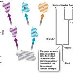
Molecular phylogeny is a method used to analyze molecular differences in heritable traits within a gene. These traits are compared to other organisms that are thought to be genetically similar on order to determine whether there is an evolutionary relationship between them. Phylogenetic trees can give us a bigger picture of the relationships between species, if they have common ancestors, or if the species or phylogenic groups can be traced back to in the phylogenic tree to a point where we can hypothesize a general time frame when one subset of a species genetically diverged from an original. It will be important to remember that a species includes organisms able to interbreed and produce viable offspring. When imagining the origins of life, we can see how putting together all evolutionary relationships would form a tree, with the most shared common ancestors at the base and trunk, and the most recent species forming the branches. Below is an example of a phylogenetic tree retrieved from Chapter 23 of Launchpad:

Looking above, one can see that the origin of the tree in question is called the “root” named so because that is the most common ancestor of species A, B, and C. the forks where we can see genetic divergence is called a node. The lines coming off of that node is called a Branch, which can be followed to yet another divergent species. The more branches observed in a phylogenetic diagram, the more speciation that occurred.

References

Biology: How life works (2nd ed.). (2017). In B. A. Berry Andrew, Biology: How life works (2nd ed.) (p. CH 23). McGraw Hill, NY: W. H. Freeman. Retrieved from http://www.macmillanhighered.com/launchpad/morris2e/4909413#/ebook/item/MODULE_bsi__F4951CED__2971__4486__BC4F__E109B2EE87D4/bsi__2ED633B9__F1E8__4C6C__82AA__0633C0C9F474?mode=Preview&toc=syllabusfilter&readOnly=False&renderIn=fne

Enzymes & Enzyme Inhibitors

Enzymes play many key roles in energy metabolism and homeostatic balance. Without them, cells would be several times more inefficient (if not nonfunctional) in its energy usage, more susceptible to foreign materials, and wouldn’t be able to utilize as many organic materials for energy. Enzymes are proteins. Like all proteins, they are made up of long chains of amino acids transcribed from DNA or RNA templates. They catalyze/accelerate the rate of chemical reactions by lowering the activation energy needed to carry out said reaction. Activation energy is the input of energy required by all chemical reactions to break/synthesize bonds for metabolism in an efficient manner. They accomplish this by stabilizing reactant(s) in their transition state, lowering the amount of free energy released to make it more available, lowering the amount of input energy needed to make or break a chemical bond. Enzymes are also energy efficient because they do not disappear after use and can continue to catalyze reactions. There are several different kinds of enzymes, and each type works on a substrate or reaction. Their specificity comes from the fact that the shape of the enzyme will determine its overall function, so therefore they are tailored to work on one substrate.

All enzymes have special folds in its structure designed to fit a specific substrate, called an Active site. Once a substrate is attached to the active site, a cofactor is normally needed in another receptor or fold of the enzyme for it to perform its function. Cofactors can include Vitamins, co-enzymes (enzymes that work in conjunction with another to form a complex), metal ions, hormones, etc.
Though most enzymes are regulated by cell processes like gene expression, hormone and neurotransmitter release (forms of cell signaling) negative feedback loop with its substrate, or cofactor availability, enzymes are greatly affected by their environment. Its shape can be altered in the presence charged molecules, very susceptible to changes in PH and light, and can be inhibited. Enzyme inhibition is a useful concept to know because inhibitors are used a lot in the medical field in drug therapy, in slowing an unwanted reaction, etc. A most well-known example is penicillin, which inhibits an enzyme used by pathogenic bacteria that repairs damage to its cell wall.

There are three types of enzymatic inhibition: Competitive inhibition, noncompetitive inhibition, and allosteric inhibition. Competitive inhibition occurs when a molecule similar in structure to the substrate binds to its active site, thus preventing it from performing its function (penicillin is an example). Noncompetitive inhibition is when an inhibitor binds to an area other than its active site. They could work by changing its shape or possibly by physically blocking the active site. Lastly, allosteric inhibitors work by adding either an activator molecule to its allosteric site (the site where activators/inhibitors bind to). Examples of allosteric regulators could be hormones, neurotransmitters, or some other type of cell signaling molecule.

References

Biology: How life works (2nd ed.). (2017). In B. A. Berry Andrew, Biology: How life works (2nd ed.) (p. CH 6). McGraw Hill, NY: W. H. Freeman. Retrieved from http://www.macmillanhighered.com/launchpad/morris2e/4909413#/ebook/item/MODULE_bsi__F4951CED__2971__4486__BC4F__E109B2EE87D4/bsi__2ED633B9__F1E8__4C6C__82AA__0633C0C9F474?mode=Preview&toc=syllabusfilter&readOnly=False&renderIn=fne

Hardy-Weinberg Principal

The Hardy-Weinberg principle is a concept conceived in 1908 by G.H Hardy and Wilhelm Weinberg. It describes a situation in which allelic or genotypic frequencies that drive evolution do not change over time in a population, and therefore that population is not evolving. The concept is based on Gregor Mendel’s law of independent assortment, which states that alleles of a trait can be separated independently from each other and can lead to fluctuations in the frequency of alleles over time. With this fact known, Hardy and Weinberg developed a series of conditions that must be met in order to determine whether or not a population is evolving. This established baseline is very useful in the study of population genetics. The conditions that must be met are as follows:

1. There can be no differences in the survival and reproductive success of individuals in the population. If a certain genotype of individuals in a population have a better chance of survival than the others or if a homozygous recessive gene expresses for some deadly condition, then we know automatically that nature will select for the genotype most fit to its environment and evolution would then occur.

2. Populations must not be added to or subtracted from by migration. If one has a population they are studying and added or subtracted individuals, the frequency of the gene(s) in question being observed would immediately change because the proportion of alleles to individuals would be thrown off. Because evolution is defined as a change in allele frequency over time in a population, a change in frequency by the addition or subtraction of that genotype would count as a population evolving.

3. There can be no mutation occurring. Simply put, mutations are one of the ways populations can evolve. It should be of note to mention that mutation is not the biggest concern because population geneticists generally study populations in a much shorter timescale than it would take for mutations to accumulate. Still, none allowed.

4. The population must be large enough to prevent sampling errors. A sampling error occurs when not enough specimens in that population are being observed. If the population is too small, then those individuals have a much bigger impact on allele frequency than one would see in an actual population and any statistical data gained could not possibly reflect the population as a whole, leading to inaccurate data. A smaller population would also be more impacted by genetic drift, which is also a form of evolution.

5. Matings within the population must be at random and not chosen or planned. If individuals with known allele frequencies were chosen to mate, It would interfere with your results, so mating must happen without any regard to phenotype, as would be the case in nature. This doesn’t affect the overall genotype frequency but not the frequency of alleles (because of independent assortment).

This test can have big applications in population genetics.On top of determining whether or not a population is/has evolved, we can also use it to make predictions on what evolutionary factors are affecting a population, predict allele frequencies in a population, and use it to look for correlations between genotype frequency and allele frequency.

References

Biology: How life works (2nd ed.). (2017). In B. A. Berry Andrew, Biology: How life works (2nd ed.) (p. CH 21). McGraw-Hill, NY: W. H. Freeman. Retrieved from http://www.macmillanhighered.com/launchpad/morris2e/4909413#/ebook/item/MODULE_bsi__F4951CED__2971__4486__BC4F__E109B2EE87D4/bsi__2ED633B9__F1E8__4C6C__82AA__0633C0C9F474?mode=Preview&toc=syllabusfilter&readOnly=False&renderIn=fne

Cytokinesis

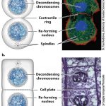
Cytokinesis is the stage in the eukaryotic cell cycle where a replicating cell divides its components into 2 separate membranes, forming daughter cells. It is widely thought that cytokinesis in eukaryotes evolved from the similar process of binary fission in Prokaryotes, yet it is a more complex process in comparison due to the nature of eukaryotic DNA replication. Cytokinesis is also more complex because eukaryotes have nuclei that must break down in the beginning of the cell cycle and reform at its end. It occurs in Both meiosis and mitosis wherein in the final stage of mitosis creates 2 diploid daughter cells with the same genetic makeup as the parent cell, and in meiosis (the reproduction of germ-line cells) it occurs twice. the first division occurs after telophase of Meiosis I, and again in Meiosis II, resulting in 4 unique haploid cells. The cells are haploid in meiosis because each daughter cell contains only one pair of chromosomes that will later recombine with another germ-cell of the opposite sex.

The Picture shown above retrieved from the Biology Launchpad website shows cytokinesis in both plants and animals. All eukaryotes perform cytokinesis to divide genetic material and cytoplasmic components amongst the daughter cells, but the mechanism used to perform the separation varies between plants, animals, and fungi. In mammals, a contractile ring made of actin filaments forms at the center of the cell, and motor proteins cause the filaments to contract and will eventually pinch the cell in two, resulting in 2 new daughter cells. Plants are different because they have cell walls and do not physically separate from each other. Once the plant cell doubles in size it will create a wall of microtubules in the center called a pragmoplast that will pull in vesicles containing cell wall components towards it during telophase. The vesicles will then fuse at the center (or cell plate) to form a cell wall that is attached to the original. Fungal cytokinesis is more like animal cytokinesis and uses a contractile ring made of actomyosin.

References

Biology: How life works (2nd ed.). (2017). In B. A. Berry Andrew, Biology: How life works (2nd ed.) (p. CH 11). McGraw-Hill, NY: W. H. Freeman. Retrieved from http://www.macmillanhighered.com/launchpad/morris2e/4909413#/ebook/item/MODULE_bsi__F4951CED__2971__4486__BC4F__E109B2EE87D4/bsi__2ED633B9__F1E8__4C6C__82AA__0633C0C9F474?mode=Preview&toc=syllabusfilter&readOnly=False&renderIn=fne

“The Mitotic Phase and the G0 Phase.” Boundless Biology Boundless, 24 Mar. 2017. Retrieved 24 Apr. 2017 from https://www.boundless.com/biology/textbooks/boundless-biology-textbook/cell-reproduction-10/the-cell-cycle-88/the-mitotic-phase-and-the-g0-phase-396-11622/

ATP

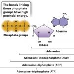
All cells require ATP to perform any action where an input of energy is needed. ATP stands for Adenosine triphosphate and is well known as the “energy currency” of all cells. We relate it to currency because ATP is not the energy itself, rather it is a molecule with a high energy value stored in its chemical bonds. Its role in metabolism is similar to the way a dollar bill represents purchasing power. The more ATP a cell contains, the more energy it has readily available for use when needed. ATP is composed of a triphosphate group (a chain of three phosphates) and adenosine (an adenine attached to a five-carbon ribose sugar). Below is a picture representing its chemical structure.

Because its energy value comes from its chemical bonds, ATP is considered a form of chemical potential energy. The reason it has such a high energy value relative to other molecules is primarily because of its unstable phosphate group. The bonds are unstable because phosphates are negatively charged and try to repel each other, but they are held together by weak hydrogen bonds. The fact that the phosphates are naturally repelling each other due to their opposing charges and are held together by weak hydrogen bonds make it easy for a cell to break the bond in a hydrolysis reaction, and in doing so will transform its stored chemical energy into mechanical energy that will be used to drive most cellular processes.
Though one molecule of ATP is structurally identical to others, the energy output from the hydrolysis of ATP can vary. The maximum output from the hydrolysis of ATP within a living cell can be up to -57kj/mol, however environmental conditions within the cell like PH and Magnesium ion (Mg2+) concentration can change this value. Note the negative sign implies energy is released from the reaction, so it is classified as exergonic. The second law of thermodynamics states that energy will be inevitably lost in the reaction in the form of heat, so it is impossible for the reaction to be 100% efficient. To compensate for this loss ATP catabolisms are coupled with chemical reactions of other forms, namely with redox reactions. The synthesis reaction of ATP in the form of a chemical equation is ADP+Pi+free energy–>ATP + H2O. ADP stands for Adenosine diphosphate and is a more stable molecule. This reaction is reversible, and therefore adding a Pi group to ADP with enough added energy will yield ATP. Clearly, without ATP, life on earth would not be possible, so understanding the role of ATP in energy metabolism is critical to understand life’s processes.

References

ATP: Adenosine Triphosphate. (2017). Retrieved April 24, 2017, from Boundless Biology Boundless: Retrieved from https://www.boundless.com/biology/textbooks/boundless-biology-textbook/metabolism-6/atp-adenosine-triphosphate-71/atp-adenosine-triphosphate-349-12938/

Biology: How life works (2nd ed.). (2017). In B. A. Berry Andrew, Biology: How life works (2nd ed.) (p.115-123, CH 6). McGraw-Hill, NY: W. H. Freeman

Final Learning Summation

Leeza-Marie Williams

Endomembrane system:

The endomembrane system consists of a group of membrane bound organelles that are found in eukaryotes. These organelles work together to modify, package, and transport lipids and proteins. It is important to know that mitochondria, chloroplasts, and peroxisomes are not included in the endomembrane system.

The endoplasmic reticulum (ER) consists of a rough ER and a smooth ER which are important in the modification process of proteins and the synthesis of lipids. Structurally, the ER consists of membranous tubules that have a hollow space called lumen, and flattened sacks.

Rough ER have ribosomes attached to its cytoplasmic surface which makes protein chains that are sent into the lumen. The protein that is not transferred fully are anchored in the membrane. Proteins inside of the membrane fold and are modified with the addition of carbohydrate side chains. Modified proteins are incorporated into the membrane of the ER, other organelles, or secreted from the cell. Proteins that are sent out of the cell are first sent to the Golgi apparatus. Phospholipids that are manufactured in the rough ER are also transported out via vesicles. Smooth ER have several functions including synthesizing carbohydrates, lipids, and steroids, detoxification, and storing calcium.

As mentioned previously, proteins that bud off of the ER are sent to the Golgi apparatus where there are sorted, tagged, packaged, and distributed to the right location. The Golgi apparatus has a cis face where it receives protein and a trans face where protein is sent away from the apparatus. A fusion occurs between transport vesicles of the ER and the cis face of the Golgi apparatus where the contents within the transport vesicles are emptied into the lumen of the Golgi apparatus. Further modification may occur to proteins and lipids within the Golgi apparatus. For example, the addition or removal of short chained molecules of sugar or phosphate groups may occur. Modified proteins are sorted based on their amino acid sequences and chemical tags and are packaged into vesicles that bud away from the Golgi apparatus at its trans face. Some of the vesicles that budded off of the Golgi apparatus are sent to the lysosome or vacuole or fuse with the plasma membrane to secrete proteins outside of the cell.

The lysosome contains digestive enzymes that breaks down structures to reuse their molecules. Lysosomes also have the ability to digest foreign particles. For example, macrophages are a class of white blood cells that folds inward to engulf pathogens. Once the pathogen is contained, it forms a phagosome when it is pinched off from the plasma membrane. The phagosome and the lysosome fuse to form a compartment consisting of digestive enzymes that destroy the pathogen. Vacuoles are an alternative to lysosomes in plants which stores water, wastes, isolates pathogens, and breaks down macromolecules.


Mitochondria:

Mitochondrion are different from most other organelles because it has its own circular DNA, similar to the DNA of prokaryotes, and reproduces independently of the cell. Therefore, mitochondrion are endosymbiotic. Mitochondria convert oxygen and nutrients into adenosine triphosphate (ATP). ATP is the chemical energy “currency” of the cell that powers the cell’s metabolic activities. This process is called aerobic respiration and is the reason animals breathe oxygen. Without mitochondria, more complex animals, like humans, would likely not exist because their cells would only be able to obtain energy with the absence of oxygen or anaerobic respiration which is less efficient than aerobic respiration.

Most mitochondria are oblong organelles between 1 and 10 micrometers in length. Both the inner and outer mitochondrial membranes resemble the plasma membrane in molecular structure. Each are 60 to 70 Ǻ thick and are composed of two layers of phospholipid molecules in between two layers of protein molecules. The outer and the inner membranes contain specific channels for molecules to transport through them. The two membranes may be connected at adhesion sites. The outer and inner mitochondrial membranes are separated from each other by a narrow space called the intermembrane space.

The outer membrane is freely permeable to most small molecules. such as ions, ATP, ADP, and nutrient molecules because it consists of transmembrane channels formed by the protein, porin. The inner membrane is freely permeable only to oxygen, carbon dioxide, and water. Its structure is highly complex, including all of the complexes of the electron transport system, the ATP synthetase complex, and transport proteins. The wrinkles, or folds within the innermembrane are organized into layers called the cristae which divides the mitochondrion into compartments running perpendicular to the long axis of the rod shaped mitochondrion. The cristae greatly increase the total surface area of the inner membrane which provides enough space for the previously mentioned structures to fit inside of the mitochondria. The matrix contains enzymes that are responsible for the citric acid cycle reactions. The matrix also contains dissolved oxygen, water, carbon dioxide, the recyclable intermediates that serve as energy shuttles. An electron carrier is a molecule that accepts an electron from one molecule and transports it into another, this movement of electrons by carriers is known as the electron transport chain. Carriers can exist in either an oxidized or reduced form, in an oxidized form the carrier is available to receive additional electrons, in a reduced form the carrier is actively carrying electrons.


Chloroplasts:

The process of photosynthesis is conducted using chloroplasts which are used to convert light energy into chemical energy. Since chloroplasts are involved with storing energy and synthesizing metabolic materials, they are categorized as a type of plastid. Various types of plastids are required depending upon the amount of light surrounds the leaf as it grows. Leucoplasts are used to synthesize starch, oil, and protein. Chromoplasts that are yellow to red synthesize carotenoids. Green chloroplasts contain chlorophyll a and chlorophyll b which are involved with absorbing light energy required for the process of photosynthesis.

To describe the structure of the chloroplast, it is enclosed in a double membrane consisting of two layers within the membrane called the intermembrane space. In terms of permeability, the outer portion of the double membrane is more permeable than the inner portion of the membrane. The stroma comprises of most of the chloroplast volume by containing a semi fluid material consisting of enzymes. Like mitochondria, chloroplast have their own DNA, ribosomes, and RNA which are found in the stroma. More complex plants have internal membranes (lamellae) with stacks (granum) of closed hollow disks (thylakoids). Lumens or internal spaces allow for the connection of thylakoids. Within the thylakoids are embedded chlorophyll molecules. Light travels in photons and are absorbed by molecules of chlorophyll. Once absorbed, electrons are emitted and hydrogen ions are propelled around the thylakoid stack. When hydrogen ions surround the thylakoid stack, formation of an electrochemical gradient occurs and the stroma produces ATP. Light independent reactions that involve carbon fixation occur in the stroma whereby low energy carbon dioxide molecules transforms into glucose, a high energy compound.


Cytoskeleton:

Eukaryotes possess three types of protein fibers in their cytoskeleton: microfilaments, intermediate filaments, and microtubules. Microfilaments are the narrowest type of protein fibers with a diameter of 7nm consisting of actin filaments which are linked monomers of protein that resembles the structure of a DNA’s double helix. Within microfilaments are actin subunits. Actin filaments have two structurally different ends that allow them to have directionality. They provide direction when myosin or motor proteins are needed to move around in the cell. Actin filaments also function as a means to enable the cell to transport protein containing vesicles and organelles. Actin and myosin work together in muscle cells to form sarcomeres which are structures of overlapping filaments. When sarcomeres slide past each other, the muscles within our body contracts. Since actin filaments have the ability to assemble and disassemble quickly, it plays a vital role in cell movement.

Intermediate filaments consist of multiple wound strands of fibrous proteins and are the in between the size of microfilaments and microtubules with a diameter of 8 -10 nm. Intermediate filaments differ than actin filaments in that intermediate filaments are more permanent and are specialized to handle tension in order to maintain the shape of the cell. They also serve to anchor the nucleus and other organelles in a specific location.

Contrary to their name, microtubules have the largest diameter at 25nm of all three types of cytoskeleton fibers. Microtubules consist of an arrangement of straw like α-tubulin and β-tubulin subunits. Microtubules are similar to actin filaments in that they have the ability to grow and shrink whenever tubulin proteins are added or removed. Since both actin filaments and microtubules have directionality, they both resist compression forces due to possessing two ends that are structurally different from one another. Without microtubules, motor proteins like kinesins and dyneins would not be able to transport vesicles within the cell. Microtubules are also important in cell division whereby microtubules assemble into spindles that pulls the chromosomes apart.


Protein trafficking:

Membrane bounded transport vesicles transport proteins embedded in the rough ER to the Golgi apparatus. The Golgi apparatus consists of cisternae which are flattened membrane sacs. Translation and modifications of proteins occurs in the rough ER. Modifications are completed in the Golgi apparatus as proteins pass through the complex to ensure that the proteins reach the correct final destination. Each region of the Golgi apparatus contains different enzymes depending on how it plans to modify proteins. As mentioned previously under the topic about the endomembrane system, proteins travel from the cis to the trans regions of the Golgi apparatus where they are then released to the lysosomes or vacuoles or outside of the cell.


Cell signaling:

There are four general categories of chemical signaling: paracrine signaling, autocrine signaling, endocrine signaling, and signaling by direct contact. The main difference between these types is the distance that the signal travels through the organism to reach the target cell.

Paracrine signaling occurs between cells when they are relatively close to one another. In paracrine signaling the cells communicate through releasing ligands that can diffuse through the space between the cells. Paracrine is critical to the process of development and to synaptic signaling of nerve cells transmitting signals.

Autocrine signaling involves a cell releasing a ligand that binds to the receptors on its own surface or inside of the cell. It is beneficial for enabling cells to reinforce their correct identities during development. Autocrine signaling is also beneficial to the spread of cancer from its original site to other parts of the body, metastasis.

The process of signals that are produced by specialized cells and released into the bloodstream where they are carried to target cells is called endocrine signaling. Endocrine signaling transmit signals over long distances. Hormones are signals that are produced in one part of the body and are carried to other targets via circulation.

Signaling by direct contact occurs when two cells bind to each other because they have complementary proteins on their surfaces. Once bound, the interaction causes the shape of one or both to change which transmits a signal. The immune system is heavily dependent upon this type of signaling whereby the immune cells use protein markers to recognize the body’s own cells from pathogens.


Endosymbiotic theory:

With regards to mitochondria, a hypothesis proposed among the scientific community, states that millions of years ago small, free-living prokaryotes were engulfed by larger prokaryotes because they were able to resist the digestive enzymes of the host organism. The two organisms developed a symbiotic relationship over time. The larger organism providing the smaller with ample nutrients and the smaller organism providing ATP molecules to the larger one. Through further evolution, the larger organism developed into the eukaryotic cell and the smaller organism developed into the mitochondrion.


Works Cited:


Caprette, David. “Structure of Mitochondria.” Rice University, 26 May    2005. Web. 9 Feb. 2017.

<http://www.ruf.rice.edu/~bioslabs/studies/mitochondria/mitotheory.html>.


Davidson, M. (2000, October 1). Chloroplasts. Retrieved April 22, 2017, from

https://micro.magnet.fsu.edu/cells/chloroplasts/chloroplasts.html


Davidson, Michael. “Mitochondria.” Molecular Expressions Cell Biology and Microscopy Structure and Function of Cells and Viruses. Florida State University, 1 Oct. 2000. Web. 9 Feb. 2017. <https://micro.magnet.fsu.edu/cells/mitochondria/mitochondria.html>.


Introduction to cell signaling. (n.d.). Retrieved April 22, 2017, from

https://www.khanacademy.org/science/biology/cell-signaling/mechanisms-of-cell-signaling/a/introduction-to-cell-signaling


McClean, P., Ph.D., & Johnson, C. (n.d.). Retrieved April 22, 2017, from

http://vcell.ndsu.nodak.edu/animations/proteintrafficking/transport_02.htm


“Structure Mitochondria.” Tutor Vista, n.d. Web. 9 Feb. 2017.           <http://www.tutorvista.com/biology/structure-mitochondria>.


The cytoskeleton. (n.d.). Retrieved April 22, 2017, from

https://www.khanacademy.org/science/biology/structure-of-a-cell/tour-of-organelles/a/the-cytoskeleton


The endomembrane system. (n.d.). Retrieved April 22, 2017, from

https://www.khanacademy.org/science/biology/structure-of-a-cell/tour-of-organelles/a/the-endomembrane-system


Electron Carriers

An electron carrier is a molecule that accepts an electron from one molecule and transports it into another, this movement of electrons by carriers is known as the electron transport chain. Carriers can exist in either an oxidized or reduced form, in an oxidized form the carrier is available to receive additional electrons, in a reduced form the carrier is actively carrying electrons. Although there are many different electron carrier, the two most common within a human body are NADH and FAD. NADH is the reduced form of NAD+ that has accepted two electrons and a hydrogen ion, furthermore, FAD is the reduced form of FADH2 that has accepted two electrons and a hydrogen ion. These electron carriers are essential in cellular respiration where they harvest electrons from glucose to produce vital ATP, redox reactions during cellular respiration add or remove electrons from the electron carriers. Cellular respiration generally occurs in three processes but can vary depending on environment and the type of cell, these processes are glycolysis, the citric acid cycle, and oxidative phosphorylation. In glycolysis, the six-carbon molecule glucose undergoes a range of chemical transformations, the electron carrier NAD+ is reduced by accepts two electrons and a hydrogen ion producing NADH.  Additionally, two molecules of pyruvate, a three-carbon organic molecule, and ATP is produced. The pyruvate molecules then enter the mitochondria where they are processed into acetyl CoA, NADH is also produced while Co2 is released. The processes then continue into the citric acid cycle, acetyl CoA is processed with a four-carbon molecule to produce ATP, NADH, and FADH2. Additionally, the four-carbon molecule is regenerated to be used again and CO2 is released. The final process of oxidative phosphorylation is now initiated, NADH and FADH2 place their electrons into the electron transport chain to become oxidized. With these electrons, the electron transport chain expels protons from the matrix to create an electrical gradient. Protons reenter the matrix with the assistance of an enzyme called ATP synthase and produce ATP, during this process the majority of ATP produced in cellular respiration is made. With the process of cellular respiration explained, it’s easy to see how vital electron carriers are in the production of ATP.

References:

https://www.khanacademy.org/test-prep/mcat/biomolecules/overview-metabolism/v/electron-carrier-molecules