Cell Structure of Mycobacterium Tuberculosis
Hello all! Here is some information regarding the cell structure for Mycobacterium tuberculosis, the cause for the horrid and contagious disease, Tuberculosis!

Mycobacterium Tuberculosis has a rod-shape and its a pretty large bacteria! The length of the bacteria usually ranges from 2 to 4 micrometers and the width is somewhere between 0.2 to 0.5 micrometers. Interestingly, Mycobacterium tuberculosis is a non-motile bacteria (Todar). Additionally, because M. tuberculosis is a prokaryotic bacteria, it is a single-celled organism.

Size and shape of M. Tuberculosis
Next, the genome of M. tuberculosis is comprised of 4, 411, 529 base pairs and 4,000 different genes. Not to mention, M. tuberculosis‘s genome has a very large guanine + cytosine content which is reflected in the amino acid content of the protein products. Mycobacterium tuberculosis is unique to other bacteria because a large chunk of its coding capacity is dedicated for the production of enzymes that are involved in lipogenesis and lipolysis. Studies have shown that lipolytic enzymes are potential biomarkers for possible diagnoses of active Tuberculosis (Badcock et al.,).

Classification of M. tuberculosis genes
Now let’s move onto the composition of the cell wall for Mycobacterium tuberculosis bacteria!
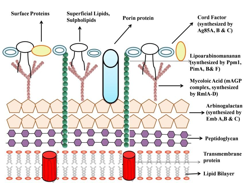
Interestingly, M. tuberculosis is not classified as either gram positive or gram negative bacteria because it does not have all the characteristics that a gram +/- bacteria would typically have; M. Tuberculosis is considered to be an acid-fast bacteria. A key factor of the M. tuberculosis cell membrane is that over 60% of it is made out of lipids. The cell wall of M. tuberculosis is unique in that it has mycolic acids, cord factor, and mAPG complex (Todar).
Mycolic acids are long fatty acids found in the cell wall of M. Tuberculosis. Because mycolic acids contribute to the waxy-like coating on the cell surface, the bacterial cells are essentially impervious to normal gram staining techniques. Hence, acid-fast staining is utilized because it can penetrate through the dense lipid layer and stain the bacterial cell; the Ziehl-Neelsen stain is used to dye M. tuberculosis with a special pink stain called carbon-fuchsin. Resultantly, the acid-fast stained bacteria will appear light pink (Todar).
Acid-fast staining technique on bacteria very similar to Mycobacterium tuberculosis!
Furthermore, cord factor is a surface glycolipid and causes M. tuberculosis to grow serpentine cords. It is evident that mostly virulent strains of M. tuberculosis possess the cord factor. The mAPG complex is essentially the cross-linkage between the peptidoglycan layer and the arabinogalactan layer; the complex provides a foundational basis for the cell wall of M. Tuberculosis and is essential for the viability of the bacteria.

M. Tuberculosis serpentine cording
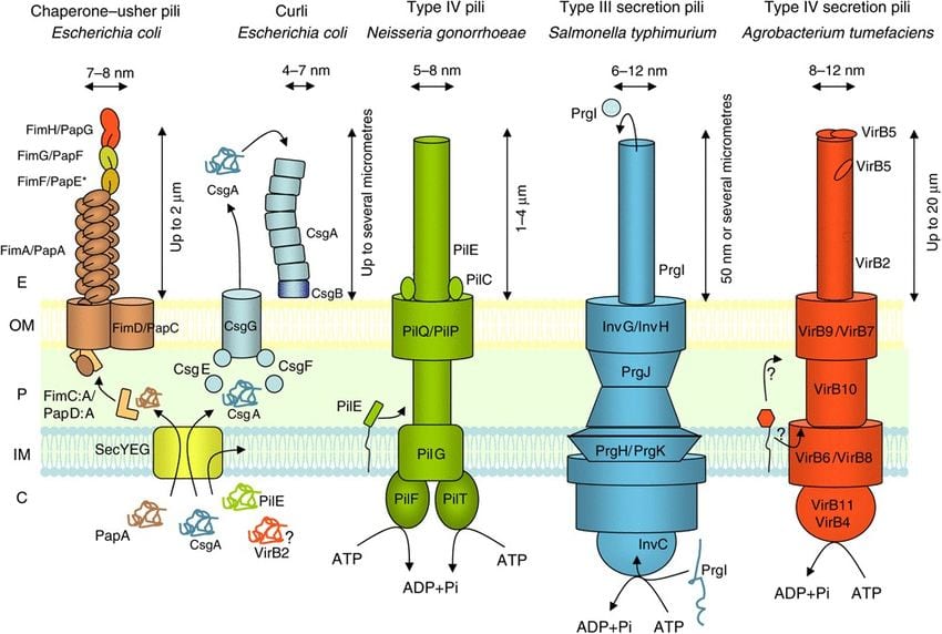
As for locomotive structures, M. tuberculosis bacteria don’t have flagella but they do have fine, aggregative, and flexible pili. Specifically, the type of pili Mycobacteria bacteria have are type IV and curli pili. Studies also show that M. tuberculosis bacteria utilize sliding motility to spread across surfaces and promote infection (Kolter et al., Pillay & Ramsugit).
And that’s it for today’s blog post! Stay tuned for next time to learn about the life cycle of M. tuberculosis!

Sources:
Badcock, K. et al., “Deciphering the Biology of Mycobacterium Tuberculosis from the Complete Genome Sequence.” Nature International Journal of Science, Springer Nature Publishing AG, 11 June 1998, www.nature.com/articles/31159.
Kolter, R, et al. “Sliding Motility in Mycobacteria.” Journal of Bacteriology, American Society for Microbiology, Dec. 1999, www.ncbi.nlm.nih.gov/pmc/articles/PMC103697/.
Pillay, M. & Ramsugit S. “Pili of Mycobacterium Tuberculosis: Current Knowledge and Future Prospects.” Archives of Microbiology, U.S. National Library of Medicine, Aug. 2015, www.ncbi.nlm.nih.gov/pubmed/25975850.
Todar, Kenneth. Mycobacterium Tuberculosis and Tuberculosis. Todar’s Online Textbook of Bacteriology, textbookofbacteriology.net/tuberculosis.html.