The project, titled “Influences of ecological niche on mechanisms of visual pathway maturation”, will be awarded $1 million over 4 years. Our goals are to determine whether the evolutionary history and ecological niche occupied by a species predicts the extent to which visual experience is required for the development of visual pathways in the brain. We will examine several rodent species, including the diurnal Chilean degu, that differ with respect to circadian activity pattern and the degree to which vision is important for behavior.
Congratulations to Precious McGowan
Precious has completed her B.S. in Biology with distinction and will be moving on to her next challenge. We are grateful for her hard work in our lab and wish her the best!
Parag Juvale joins the lab!
Parag is a 2nd year M.S. student in the Biology department. His background is in the use of natural compounds to fight cancer. His experience with Western blotting will be of great benefit to our progress. He will be working on signaling molecules that are important both for axon guidance during brain development, and for vascularization of growing tumors.
Jenny Kim joins the lab!
Jenny is an undergrad in the Neuroscience program and will be working on a visual perception assay in the lab as well as helping us out with histology and the mice. She has already made herself indispensable and we expect great things from her in the future.
Pallas Lab wins a seed grant!
Thanks to the Brains and Behavior Program for supporting our work.
A Most Amazing Paper in Neuroscience!
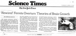
A 2000 Nature paper from the Sur lab at MIT, coauthored by Sarah Pallas, again made the list of Most Amazing Papers in Neuroscience! This perceptual behavior study, initiated by Dr. Pallas while she was a postdoc in Sur’s lab, and subsequently completed by another postdoc, Laurie von Melchner, answered a long-standing question about “labeled lines” in sensory pathways and the limits of experience-dependent plasticity. It had been thought that sensory brain areas were intrinsically labeled with a specific sensory modality; that is, activation of auditory cortex should always produce the sensation of sound, regardless of how that activation is achieved. For example, if you (carefully!) push on your closed eyelid with your finger, you will see light flashes. We discovered, however, that ferrets whose retinal axons had been rewired during development to invade the auditory pathway could perceive light using their auditory cortex. These surprising results demonstrated that sensory experience and perceptual learning can strongly influence the wiring of sensory cortical circuits, even to the extent that perceptual assignments of cortical regions can be switched between modalities. They also point out that the neural circuits across different cortical areas are actually very similar, and serve to tell us about our world using whatever inputs are available. Subsequent studies by Yuting Mao in the Pallas lab demonstrated that this flexibility is not without cost; residual auditory function in the rewired auditory cortex is compromised. Nonetheless, the implications of these findings for recovery of brain function after traumatic brain injury or stroke are quite exciting. They also provide an excellent example of how flexibility and plasticity in neural circuits can facilitate an adaptive response to evolutionary changes, such as mutations that affect the size or function of sensory pathways. For example, during primate evolution, the relative size of cerebral cortex compared to the brain as a whole has expanded considerably, resulting in a cerebral cortex that is enlarged and more convoluted in humans than in our primate ancestors. This extra cortical tissue has been invaded by existing sensory structures through competition for target space and experience-dependent plasticity, providing additional circuits to devote to processing that sensory input and thus a survival advantage. If such a mutation also affected the germ cells, it would be inherited by the descendants, increasing the frequency of this genetic variation in the population.
See:
- Harrington IA, Grisham W, Brasier DJ, Gallagher SP, Gizerian SS, Gordon RG, Hagenauer MH, Linden ML, Lom B, Olivo R, Sandstrom NJ, Stough S, Vilinsky I, Wiest MC. (2015) An Instructor’s Guide to (Some of) the Most Amazing Papers in Neuroscience. J. Undergrad. Neurosci. Educ. 14:R3-R14.
- Mao YT, Pallas SL. (2012) Compromise of auditory cortical tuning and topography after cross-modal invasion by visual inputs. J Neurosci. 32:10338-51.
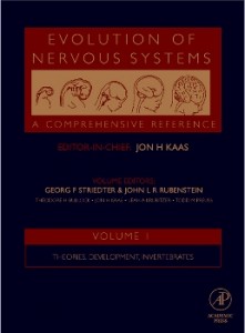
- Pallas S.L. (2007) Compensatory innervation in development and evolution. In: J. Kaas (ed.), Evolution of Nervous Systems, Vol. 1, G.F. Striedter and J.L.R. Rubenstein (eds.): Theories, Development, and Invertebrates, pp 153-168. Elsevier Academic Press, Amsterdam.
- Swindale NV. (2000) Brain development: Lightning is always seen, thunder always heard. Curr Biol. 10:R569-71.
- von Melchner L, Pallas SL, Sur M. (2000) Visual behaviour mediated by retinal projections directed to the auditory pathway. Nature. 404:871-6.
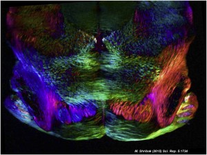
Amazing images: polychromatic polarization microscopy of Tim Balmer’s brain slice preps
Michael Shribak of MBL, and Tim Balmer, postdoc at OHSU’s Vollum Institute, 2012 Grass Fellow and former Pallas Lab member, collaborated to produce these beautiful images of unstained, transparent mouse brain slices. The paper describing the polychromatic polarization microscopy method that yielded these colorful images was recently published at Nature.com/ Scientific Reports.
Balmer, T. S. & Pallas S. L. (2015) Visual experience prevents dysregulation of GABAB receptor-dependent short-term depression in adult superior colliculus. J. Neurophysiol. 113, 2049–61.
Shribak, M. (2015) Polychromatic polarization microscope: bringing colors to a colorless world. Nature Sci. Reports 5: 17340.
AAAS CEO Rush Holt comes to Atlanta to seek input in Town Hall
Holt takes a question from Professor Pallas about helping teachers
teach accurate evolutionary biology concepts.
Annual meeting, Society for Neuroscience
Congrats to David Mudd on his Brains & Behavior Fellowship award
- 2 of 3
- « Previous
- 1
- 2
- 3
- Next »


