The following instruments require training
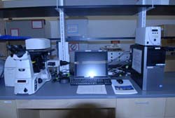
Arcturus XT Micro Dissection System with Nikon Eclipse Ti Microscope
Arcturus Microdissection System 921
Microgenomic technologies provide the tools necessary to examine expression profiles from samples as limited as a single cell. Laser capture micro-dissection (LCM) is an advanced separation technology that enables the isolation of desired pure cell populations from heterogeneous tissue samples. LCM utilizes an infrared laser pulse system which adheres cells of interest to a transparent thermoplastic film, preserving essential cellular and morphological characteristics, while maintaining the integrity of biomolecules such as DNA, RNA, and proteins. UV laser cutting may also be utilized in conjunction with LCM allowing for the rapid isolation of larger populations of cells.
Laser capture (5-10 micron section) UV cutting laser (up to 50 micron thickness) Isolate cells for DNA/RNA and Protein extraction.
Located in PSC 871. Please contact Mary Karom at mkarom@gsu.edu or 404 413 6310 for information and training.
ECHO MRI 700 BODY COMPOSITION ANALYZER

ECHO-MRI-700
EchoMRI-700 Whole Body Analyzer – The EchoMRI™700 Analyzer deliver precise body composition measurements of fat, lean, free water, and total water masses in live animals weighing up to 700 grams respectively. Small Animals – mice, rats, hamsters, within the weight range.
Scanning takes 0.5 – 3.2 minutes, depending on the precision options.
The live animals need no anesthesia and no special preparation before measurement. Approved animal protocols are necessary prior to use.
EchoMRI™ Analyzers are exceedingly easy to operate. A training period of one hour is sufficient for individuals without prior knowledge.
Numeric results are stored and can be viewed again later at any time, as well as extracted either into Excel and ASCII files or into Access database.
The EchoMRI 700 Analyzers are stand-alone units, on 4 casters for easy relocation. They use standard power sources, and are completely shielded and safe to run.
Does not produce pictures or images
Located in PSC 830

TriCarb 2910 (black top middle wall)
TriCarb 2910 TR Liquid Scintillation Counter
Use TRI-CARB Scintillation Counter – Training required and Approved Radiation Protocol
Benchtop liquid scintillation analyzer for detecting small amounts of alpha, beta, and gamma radioactivity. The scintillator converts ionizing radiation for teh radionuclide into photons of light measuring photo intensity.
Energy Range: 0-2000 keV. Check spec sheets recieved with your isotope for this range.
Sample of nuclides in library measured: Tritium H3, Carbon C14, Iron Fe 55 titrated to decrease specific activity, Sulfur S35, and Phosophorus P32 and P33. Contact regarding other isotopes to add to the library. Vial size 20 mls.
Located in PSC 921
iMARK ABSORBANCE MICROPLATE READER

iMark spectrophotometer plate reader
Filters 405nm, 415nm, 450nm, 490nm, 595nm, 655nm and 750nm
The iMark Absorbance Plate Reader is an eight-channel, vertical pathlength photometer
- Measures the absorbance of the contents in a 96 well microtitration plate
- It can perform single or dual wavelength measurement
- Reports absorbance values to three decimal places
- Custom filters between 400-750nm may be ordered from www.bio-rad.com
- Located in PSC 921
PARAFFIN WAX HISTOLOGY EQUIPMENT
Paraffin Infiltration, Embedding and Sectioning
Paraffin embedding is used to prepare the tissue specimen so it does not shred upon slicing. The tissue specimen is infused with paraffin (wax) and placed within a mold to which more melted paraffin is added. The result is a small cube of wax with the paraffin-filled tissue specimen within it. Tissue embedding is routinely used to prepare tissue biopsies for examination.
The tissue embedding process involves several steps.
-

- Step 1 – Infiltration
-

- Step 2 – Embedding
-

- Step 3 – Rotary Microtome Paraffin
1. Infiltration – STP 120 – on a table beside the Embedding system (Histostar)
2 Embedding – Histostar 4
3. Sectioning – Finesse Rotary Microtome, water bath and slide warmer
A protocol is necessary for this step of tissue processing. The process includes fixation, dehydration, clearing agents and paraffin wax. In some cases fixation is performed prior and the process starts with dehydration. Type of tissue and downstream applications determine protocol.
- A programmable, carousel tissue processor
- Tissue samples are placed in cassettes which are placed in an organizer tissue basket
- The tissue basket is placed on the operating head assembly which lowers into the containers below.
- There are 7 reagent (alcohols or fixative) containers, 3 metal clearing agents (Citrosolv is used in core only), and 2 wax baths.
- Located in PSC 863/841 middle of the room beside the Histostar 4
- Rotary Microtome, water bath and slide warmer are in PSC 921

Autosharp 5
SHANDON AUTOSHARP 5
- Automatic Sharpener for steel microtome knives.
- The steel knife is clamped onto a suitable holder which is then fitted onto an arbor mounted above a special lapping plate.
- The knife is sharpened on a rotating lapping plate. C-Type and D-Type knives.
- Located in PSC 753 – Contact Mary Karom for training

Genysis 5
GENESYS 5 SPECTROPHOTOMETER
- UV wavelength
- Sample is placed in cuvettes for measurement
- Measurement of DNA, RNA and Oligos
- Measure Protein assays such as Bradford and Lowery
- Located in 921 – Contact Mary Karom for training

NanoVue
GE NANOVUE PLUS SPECTROPHOTOMETER
- Measurement of nucleic acid and protein samples. Samples of 0.5 uL to 2 uL can be pipetted and then recovered from plate.
- Protein 4 uL is suggested volume and can be recovered from plate.
- Wavelength 200-1100: calibration automatic upon switch on
- Wavelength accuracy +/- 2nm, reproducibility +/-0.5nm
- Located in PSC 921 – Contact Mary Karom for training
CLARITY TISSUE/WHOLE BRAIN IMMUNO STAINING SYSTEM
Smart Clear II Pro and Smart Label Pro by Life Canvas Technologies, please see web site www.lifecanvastechnologies.com for information. This requires a Sheet-Light Microscope for imaging upon completion, there is one available in NSC contact Mary Karom for information.
Located in PSC, 8th floor Aras Petrulis Lab