Well hello again!
The microbe is so very small
You cannot make him out at all
But many sanguine people hope
To view him under a microscope
His jointed tongue which lies beneath
A hundred curious rows of teeth
His seven tufted tails with lots
Of lovely pink and purple spots
Upon each of which a pattern stands
Composed of 40 separate bands
His eyebrows are a tender green
All these things have never yet been seen!
But Scientists, who ought to know
Assure us that it must be so!
Oh, let us never never doubt
What nobody is sure about!
-Hilaire Belloc
First we talked about the relative SCALE of microorganisms… puts it all into perspective! Why does size even matter for a cell? What are some downsides to being very small or very large for a bacteria?
Bacteria also come in a variety of shapes. If the default shape is spherical, why do some of them go out of their way to be something other than spherical?
We talked briefly about cellular components, although this is something covered by your homework (you did your homework, right?)
We talked membranes and why they are important for bacteria. Is there something, maybe, kind of, critically important in a bacterial membrane that a eukaryote DOESN’T have in its membrane?
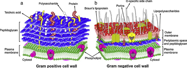
Next we identified the differences between Gram positive and gram negative cells.
https://microbewiki.kenyon.edu/index.php/Efficacy_of_antiseptic_treatments
Peptidoglycan was next on the menu: monomer structure and the purpose of the PTG. How is the PTG layer made? Why do antibiotics work against it? Here’s the animation we used in class.
Next was a brief discussion of the possible outer layers (spoiler alert, not all bacteria have them!) and what they are made of.
The flagella was next, how they can be arranged and how the swimming they facilitate is different than the twitching or gliding motion caused by other types of movement features.
Swimming:
Ooozy slimy movement:
Twitchy bacteria (pili)
And finally, the INTERNAL flagellum of spriochetes, which makes them move in a corkscrew like fashion (and really well through sludge!)
We learned about how spores are formed and how they let bacteria last through pretty much everything including space travel and the apocalypse.
And we finished up with biofilms, the slimy nasty communities that bacteria engage in to protect themselves from the outside world and facilitate their communication (through quorum sensing? Stay tuned for next week!)
******short break******
“Somewhere, something incredible is waiting to be known”
Carl Sagan (who wrote “the pale blue dot”)
We talked more in detail about the different types of stains, including the Gram stain, and why the decolorizing step seems to be the key feature of the process.
Gram positive cells staining PURPLE and Gram Negative cells staining PINK.
We then went through a litany of microscope types, including the light microscopes (bright field, dark field, confocal, phase contrast, fluorescent) and the non-light microscopes (electron, Atomic Force, and X-ray). There was a discussion of MAGNIFICATION versus RESOLUTION and how to improve both. Here’s a really nice website with more information about microscopy.
So, here are the takeaway important concepts of this week (and your study guide!)
- The differences between prokaryotic and eukaryotic cells
- The differences between Gram + and Gram – cell walls
- Draw the peptidoglycan monomer and how they connect to other monomers
- Can you describe why bacteria die when exposed to an antibiotic that inhibits peptidoglycan?
- What are the different types of bacterial motion and what structures are associated with each?
- A conceptual question…. why is the structure of the peptidoglycan layer (like a chain link fence) ideally suited to bacterial life? Why this type of structure and not something more solid like a concrete wall?
- Describe why stains are used
- Understand the process of the Gram stain and how it differentiates Gram + and Gram – cells
- Be able to determine what type of microscope would be best for a given job, what does each of them do best?
- What is numerical aperture and how is it calculated?
- What is a biofilm made from and why is it constructed in the first place?
See you next time!
Questions or comments about this content? Leave’em below!