April 25

The History of Malaria in the US

While the history of the CDC begins with malaria, the history of malaria begins far earlier, as far back as ancient Mesopotamia and Egypt. Based upon writings from these ancient cultures describing periodic fevers characteristic of malaria along with the detection of the malaria antigen from ancient Egyptian remains, it can be concluded that malaria has been around since long before Western civilization, much less the United States. Historical accounts make it clear that malaria found its way into diverse groups of people, eventually spreading through most of Europe, most likely facilitated in part by Roman expansion. Population expansion in the subtropical climates of India and Southeast Asia led to increased habitation of wetlands as opposed to the dry Indus River Valley. This further strengthened the footing of malaria as a parasite of human peoples around the Eurasian continent, as well as Africa.

    Malaria soon found its way to the Americas as an unwanted passenger of exploratory expeditions by conquistadors and eventually colonists of the new world. This meagre introduction of malaria was soon followed by a far larger importation at the hands of the Atlantic slave trade. Africans infected with malarial pathogens, both Plasmodium vivax and Plasmodium falciparum, were brought to the new world as slaves. They were seemingly healthy due to adapted immunity in the form of sickle cell anemia or G6PD deficiency, but were in fact hosts to the deadly pathogen. By the time that the United States had declared themselves independent, malaria had already taken hold in any suitable climate (tropical) in the United States and Latin America. Malaria plagued the United States throughout the 19th century, spreading from coast to coast on the backs of those exercising manifest destiny and causing the deaths of untold numbers of Native Americans in the process.

Efforts were made soon after the turn of the 20th century to control malaria in the United States as well as U.S. occupied Cuba and the areas of construction for the Panama Canal. After the request and reception of funds to the U.S. Public Health Services from congress to fight malaria in 1914, control activities were established around military bases in regions of the United States which seemed particularly “malarious”. While these early steps improved conditions around military bases, it was in 1933 that the Tennessee Valley Authority collaborated with the United States Public Health Services to combat the disease amongst all populations, military and civilian alike. The ability for mosquitoes to breed, and thus spread the disease, was curtailed dramatically by controlling water levels in known mosquito breeding grounds and applying insecticide in all surrounding areas. Through these efforts malaria was, for all intents and purposes, eliminated from the United States by 1947.

    Malaria did not go down without a fight in the early 1940’s, however. As World War II fell into full swing malaria proved to be even more deadly than the enemy forces in the beginning of the campaign in the pacific. Malaria Control in War Areas (MWCA) was founded in order to do essentially what the United States Public Health Services had done in 1914: control malaria around military training sites and bases in the Southern United States. MWCA also sought to inform local health officials about methods and strategies to use in the control of malaria, so that they could continue after the war. In 1946 the MWCA gave birth to an offshoot in the form of the Communicable Disease Center (CDC), which immediately began working on further controlling and combating malaria in the United States. The CDC, along with health agencies from 13 southern states, formed the National Malaria Eradication Program in 1947 and made quick strides in its mission to eliminate malaria. Four years and over 4.6 million housespray applications later, malaria was officially considered eliminated from the United States in 1951.


Citations


“A Brief History of Malaria.” Edited by Gelband H, Saving Lives, Buying Time: Economics of Malaria Drugs in an Age of Resistance., U.S. National Library of Medicine, 1 Jan. 1970, www.ncbi.nlm.nih.gov/books/NBK215638/.


“CDC – Malaria – About Malaria – History.” Centers for Disease Control and Prevention, Centers for Disease Control and Prevention, 14 Nov. 2018, www.cdc.gov/malaria/about/history/index.html.

April 20

The Life Cycle of Plasmodium

The life cycle of Plasmodium takes place in two distinct environments, a human host and a female Anopheles mosquito. 

Anopheles stephensi.jpeg

Anopheles mosquito

As shown in the graphic below, the life cycle of Plasmodium begins with the injection of sporozites from the mosquito’s salivary glands while the mosquito takes a blood meal from the human host. It then infects a host liver cell, where it forms a schizont that will then rupture, releasing merozites  which will then begin to infect the red blood cells.

Image result for plasmodium merozoite

Plasmodium Merozite

Once the blood cell has been infected, the ring structure known as a trophozite is formed. From here the trophozite can either mature and form another schizont (asexual reproduction), or it can differentiate in to gametocytes.

Figure C: Schizont of <em>P. falciparum</em> in a thin blood smear. Trophozoites are also seen in this image.”></p>
<p style=Schizont

From here a blood parasite (the female Anopheles mosquito) is required to ingest the male and female gametocytes (termed microgametes and macrogamtes respectively) for the life cycle to continue. Once ingested and brought to the mosquito’s stomach, the gametocytes begin the sporogonic cycle.

Figure A: Gametocyte of <em>P. falciparum</em> in a thick blood smear. Note also the presence of many ring-form trophozoites.”></p>
<p style=Gametocytes in thick blood smear

During this cycle the male (micro) gametes penetrate the female (macro) gametes, forming a zygote (sexual reproduction). The zygotyes develop in to ookinetes which are an elongated, motile form of the Plasmodium. These ookinetes then move to the gut wall of the Anopheles mosquito, where they mature and develop in to their final form, the oocysts. Finally, the cycle is completed by these oocysts rupturing to release sporozoites which move to the salivary glands so that they may infect a human host upon the next blood meal.

Graphic of Plasmodium Life Cycle


Plasmodium is heterotrophic, obtaining its energy requirements from its host cells. Plasmodium does not require special nutrition to carry out its life cycle, only specific environments.

Two informative videos on the life cycle of Plasmodium!


April 11

Plasmodium Cell Structure

Plasmodium as a genus are small protozoans that exist as unicellular organisms. For an example of their size, the species P. falciparum are on average “1/5 the diameter of a red blood cell” , making them approximately 1.44 µm in diameter. P. falciparum has been found to have a genome consisting of 14 chromosomes with a total of 5369 genes.

Figure A: Rings of <em>P. falciparum</em> in a thick blood smear.”></p>
<p style=
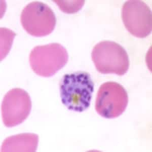
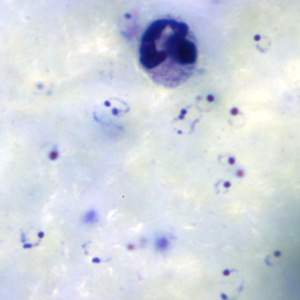
Rings of P. falciparum in a thick blood smear

Plasmodium are not bacteria and as such do not have a uniform gram stain pattern, for example P. vivax is known to display a gram positive or gram negative stain depending on the conditions. Plasmodium have a fairly typical eukaryote cell membrane, but also have several surface structures including rhoptries containing parasite proteins and micronemes that contain specialized proteins used for adhesion to host cell bodies as well as for motility. These surface structures are actually semi protruding organelles.Figure E: Rings of <em>P. falciparum</em> in a thick blood smear.”></p>
<p style=
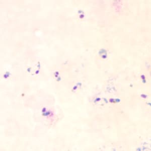
Rings of P. faciparum in a thick blood smear

Plasmodium is not motile in all of its life stages, but when it is it relies on external actin filaments to do perform taxis.

 Figure G: Rings of <em>P. falciparum</em> in a thin blood smear.”></p>
<p style=
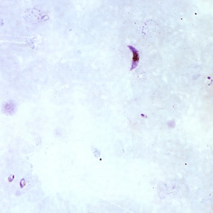
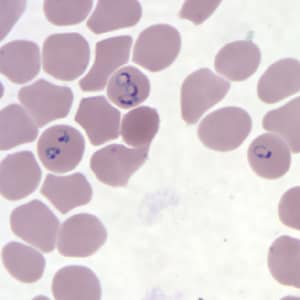
Rings of P. falciparum in a thin blood smear


Images of P. falciparum found here


A cool video of P. Falciparum invading a red blood cell!

April 3

A Trip to the CDC Museum

For my site visit I decided to go to the CDC museum as it was conveniently located, free to attend, and offered a rather deserve selection of microbes from which to choose. While at the museum I slowly meandered through the three levels of exhibits (spending a tad too much time ogling the electron microscope from the 30’s) and learned about many diseases which the CDC has sought to eliminate or at the very least reduce their rates of propagation and mortality. I was particularly take by three displays: a rather expansive timeline detailing the CDC’s efforts during the AIDS epidemic, beginning with determining what the disease even was and thus what to call it, a smaller display about viewing violence through the lens of epidemiology along with a case study in which the CDC did exactly that, and finally a detailed display about malaria and its ties to the origin of the CDC itself. I was surprised to learn that at a time after World War II, malaria was actually present and problematic in the United States, even being the disease that lead to the creation of the Public Health Service’s “Malaria Control in War Areas” which would go on to become the CDC. Until viewing this display I had been wholly ignorant not only in the role malaria played in the founding of the CDC, but that malaria had ever been an issue in the continental United States at all. This is what made me decide to make my blog about malaria and its causative agent, Plasmodium. As funny as it sounds, I was excited to learn how little I new about something so commonly talked talk about, and that I thought I essentially “had pegged” as what it was and where it was a major issue, as well as learning that it played such and important role in the creation of the Centers for Disease Control, which is a powerful force for good in modern times. The site did not have images of plasmodium itself (I have since done outside research to fix this), but did have several interesting historical images include a map of affected areas, pro-pesticide propaganda posters, and even an antique insecticide sprayer!