Last week I introduced my microbe, Mycobacterium tuberculosis. This week we’re going to dig a little deeper and examine this bacterium further.
Some fast facts on M. tuberculosis:
i. Morphology

http://textbookofbacteriology.net/tuberculosis.html
It’s a bacillus meaning it has a rod shape. In terms of size, it’s typically 2-4 µm in length and 0.2-0.5 µm in width. It is a single-celled organism that can live in colonies.
ii. Genome
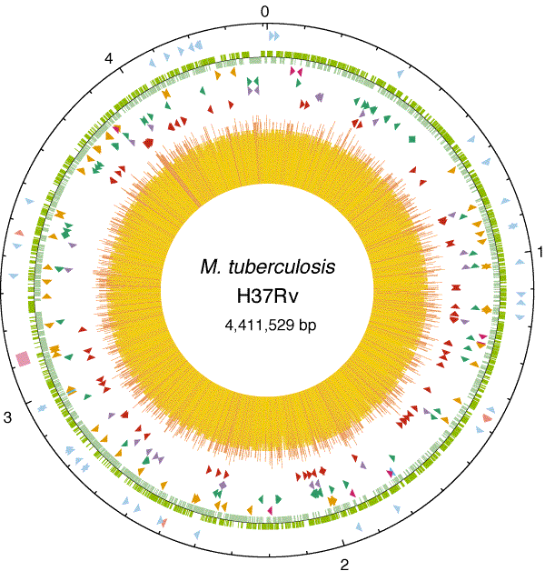
 Circular map showing chromosome of M. tuberculosis
https://www.nature.com/articles/31159
The specific strain H37Rv, of M. tuberculosis, is 4,411,529 base pairs and about 4,000 genes. It’s interesting because compared to other bacteria, a large portion of the genome is dedicated to enzyme production for the anabolism and catabolism of fats.
iii. Structure
This bacteria is neither gram (+) nor gram (-). It posses a unique cell wall that contains peptidoglycan but is comprised of mostly lipids. The lipids being: mycolic acids (for protection), cord factor (for toxin to mammals), wax-D (for cell envelope). Lipids contribute to cell’s resistance to antibiotics, lysis, and macrophages. There are no locomotive structures because the cell is non-motile.
It’s interesting to see how this bacterium compares and contrasts to typical bacteria. Next week, when we look at metabolism and life cycle, these differences may help explain how this bacterium gained notoriety for being so resistant and deadly.
Ah, quite the cliffhanger there, I can only assume that learning about the metabolism and life cycle of the bacteria will explain why such a large portion of its genome is dedicated to enzymes which catalyze lipid catabolism. As for why lipid anabolism takes up such a large chunk of the genome, I would infer that this is because the cell wall is composed of and dependent upon lipids.
It is interesting that Mycobacterium tuberculosis is neither a gram negative or gram positive and I think it is due to the cell wall. Since the cell wall is mainly lipids, it made the cell hard to be able to stain. I found out that the Ziehl – Neelsen can be used on the microbe to stain it with carbol- fuchsin resulting in a pink bacterium.